
Actinic Keratosis
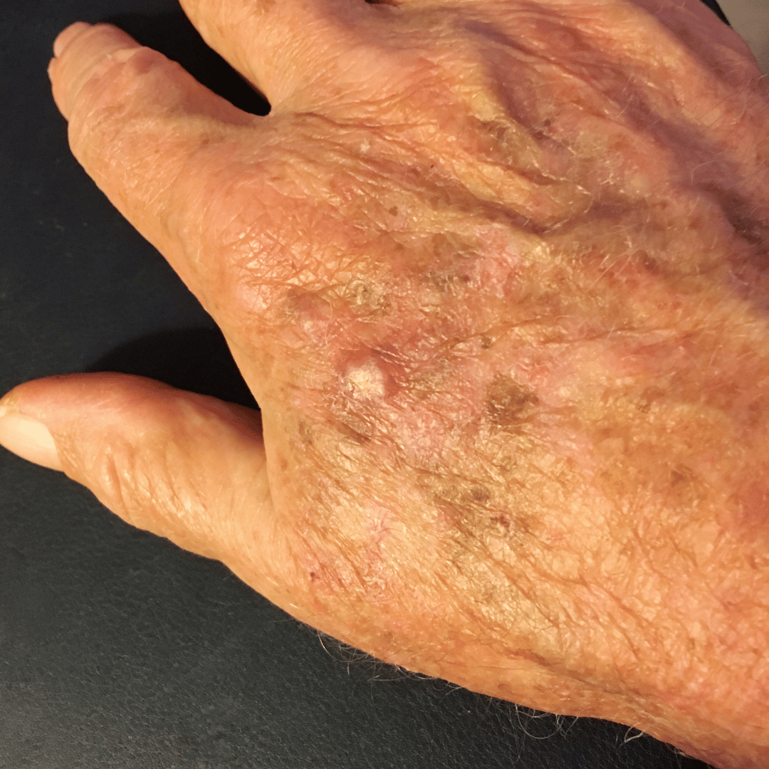
We address both visible lesions and the wider field of sun-damaged skin, using a combination of targeted and field-based approaches to reduce progression risk and support healthier skin architecture over time.

MEDICAL GROUP
The Spierings Medical Group provides specialist assessment and management of actinic keratosis, offering consultant-led care for patients with precancerous lesions resulting from cumulative UV damage. While actinic keratoses are not malignant in themselves, a proportion may progress to squamous cell carcinoma if untreated, making early identification and appropriate management clinically important.
These lesions develop when keratinocytes, the predominant cells in the epidermis, undergo DNA damage due to chronic sun exposure. Over time, this damage leads to abnormal cell proliferation and the formation of clinically visible lesions.
Although actinic keratoses are not malignant in themselves, they are considered part of a spectrum of early skin cancer development. A proportion may progress to squamous cell carcinoma if left untreated, making early identification and management clinically important.
What are the Types of Actinic Keratosis?
Actinic keratoses can present in a variety of clinical forms, often reflecting the degree and depth of cellular atypia.
- Classical (Erythematous) Actinic Keratosis - Typically presents as a rough, scaly patch with a red or pink base. Lesions are often more easily felt than seen, with a characteristic sandpaper texture.
- Hypertrophic Actinic Keratosis - A thicker, more elevated form with a pronounced keratin build-up. These lesions may resemble warts and can be more resistant to treatment.
- Atrophic Actinic Keratosis - Appears as a flatter, less scaly lesion with subtle surface change, often requiring closer clinical examination for detection.
- Pigmented Actinic Keratosis - Contains increased melanin, giving a darker appearance. It may mimic other pigmented lesions, necessitating careful differentiation.
- Actinic Cheilitis - A variant affecting the lips, most commonly the lower lip. It presents with dryness, scaling, and loss of the normal lip border definition, and carries a higher risk of progression if untreated.
What Causes Actinic Keratosis?
Actinic keratosis is the result of long-term ultraviolet radiation exposure leading to cumulative DNA damage within epidermal cells. UV radiation induces mutations in key regulatory genes responsible for cell growth and repair. Over time, these mutations impair normal cellular turnover, allowing atypical keratinocytes to persist and accumulate.
The concept of field cancerisation is central to understanding AK. Rather than isolated lesions, the surrounding skin has often undergone widespread subclinical damage, explaining why multiple lesions may develop within the same area.
Individuals with lighter skin types, a history of significant sun exposure, or advancing age are at increased risk due to reduced natural photoprotection and cumulative exposure over time.
How is Actinic Keratosis Treated?
Management is directed at both visible lesions and the wider field of sun-damaged skin, with the aim of reducing progression risk and restoring healthier skin architecture.
- Lesion-Directed Therapy - Targeted treatments focus on individual lesions
- Cryotherapy (controlled freezing) to destroy abnormal cells
- Curettage for thicker or resistant lesions
- Field Therapy - Given the presence of subclinical damage, broader treatment approaches are often required:
- Topical agents that selectively target atypical cells
- Photodynamic therapy (PDT), which uses light-activated compounds to treat wider areas of affected skin
- Adjunctive Skincare and Barrier Support - Restoration of the skin barrier plays a supportive role in improving overall skin resilience and tolerance to treatment.
Actinic keratosis represents an early stage within the continuum of UV-induced skin carcinogenesis. While individual lesions may remain stable, others carry the potential for progression. Regular skin assessment is therefore essential, particularly in individuals with significant sun exposure history. Early detection allows for timely intervention and reduces the likelihood of more advanced disease.
Consistent photoprotection is fundamental in both prevention and management. Daily use of broad-spectrum sunscreen, combined with protective clothing and behavioural sun avoidance, reduces further DNA damage and limits the development of new lesions. In high UV environments, such as the Middle East, ongoing sun protection is particularly critical in maintaining long-term skin health.

Choosingtherightdoctororclinicshouldn'tfeelrushed.
If you have a question or want to know more before booking, just ask!