
Facial Moles
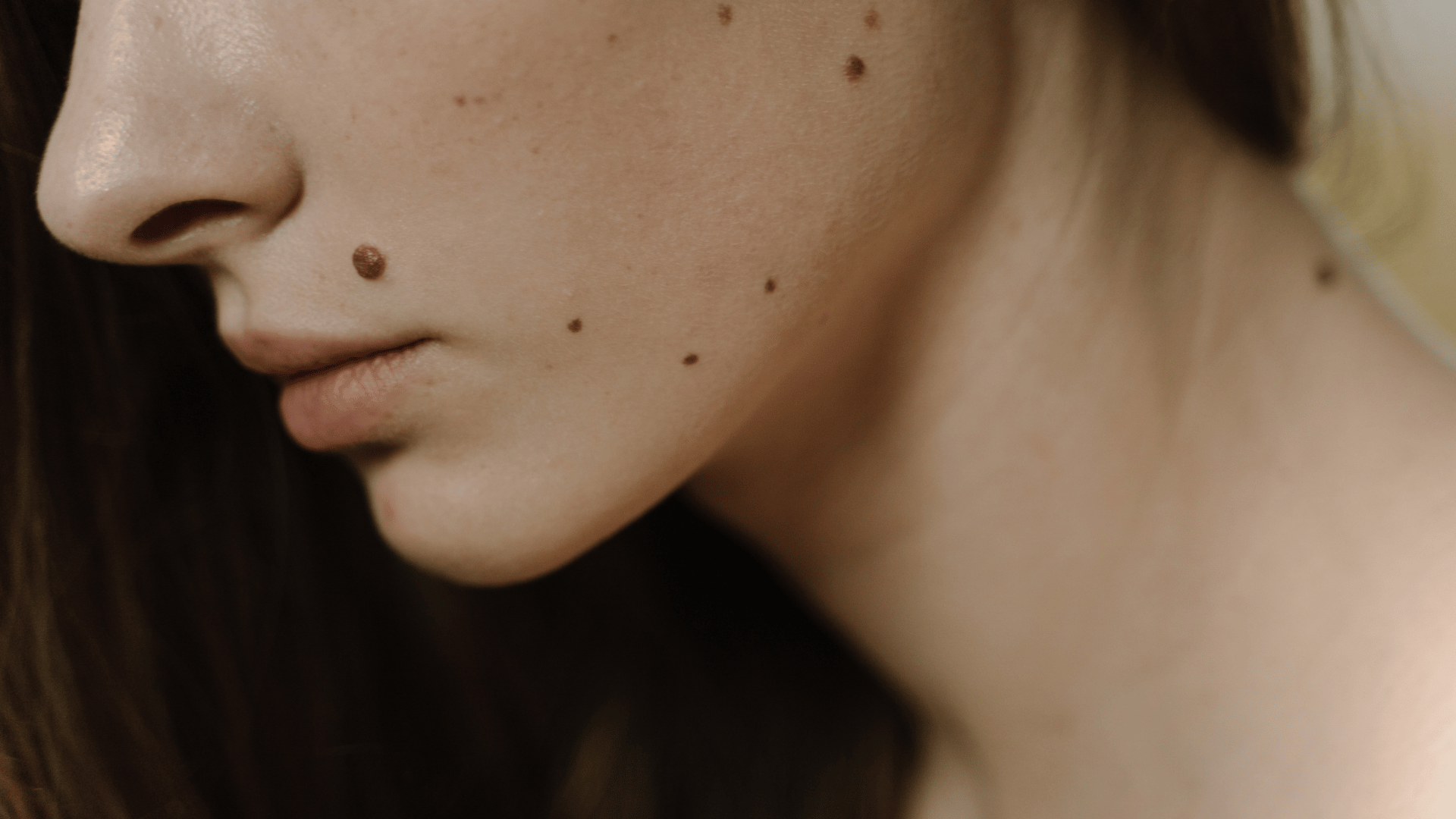
Professional monitoring is a key pillar of long-term skin health. We assess each mole carefully, explain what we find in clear language, and recommend the most appropriate course of action — whether that is reassurance, structured surveillance, or surgical excision.

MEDICAL GROUP
The Spierings Medical Group provides specialist mole assessment and monitoring, offering consultant-led dermoscopy and structured surveillance for patients with moles that have changed, concern them, or require regular review. Moles are among the most common benign skin lesions, and the overwhelming majority are entirely harmless. However, changes in their appearance can occasionally signal something that warrants clinical attention.
Melanocytic Naevi — What Your Skin Is Telling You
Moles — clinically termed melanocytic naevi — are among the most common benign skin lesions. They arise when melanocytes, the skin's pigment-producing cells, cluster together instead of distributing evenly across the epidermis. The result is a localised concentration of pigment that can appear anywhere on the body, in a spectrum of colours, sizes, shapes, and textures.
Most emerge during childhood and early adulthood and remain morphologically stable throughout life. While the overwhelming majority are entirely harmless, changes in their appearance can occasionally signal something that warrants clinical attention — making professional monitoring a key pillar of long-term skin health.
What Causes Moles to Develop?
The tendency to develop moles is strongly genetically determined. Beyond heredity, several environmental and physiological factors contribute:
- Genetic predisposition — a family history of multiple naevi significantly increases likelihood
- Ultraviolet radiation — sun exposure, particularly during childhood, directly promotes melanocyte proliferation
- Hormonal fluctuations — puberty and pregnancy can trigger the appearance or darkening of existing moles
- Skin phototype — fairer skin types tend to accumulate more moles with cumulative UV exposure
Cumulative UV exposure does not merely increase mole count — it also raises the risk of atypical mole development and, in susceptible individuals, melanoma.
Clinical Types of Moles
- Junctional Naevi: Flat, uniformly pigmented lesions at the dermo-epidermal junction. They typically appear as smooth, well-defined brown or dark spots — the most classic mole presentation.
- Compound Naevi: Slightly elevated moles containing melanocytes in both the superficial and deeper skin layers. Often textured and may contain terminal hair follicles.
- Intradermal Naevi: Raised, flesh-coloured or lightly pigmented lesions common in adults, where melanocytes have migrated fully into the dermis. Typically soft and dome-shaped.
- Atypical (Dysplastic) Naevi: Moles exhibiting irregular features including colour variation, indistinct borders, and asymmetry. Benign, but associated with elevated melanoma risk and requiring structured surveillance.
- Congenital Naevi: Present at birth. Large congenital naevi carry a higher lifetime risk of malignant transformation and require ongoing specialist monitoring.
The ABCDE Warning Signs — When to Seek Advice
The following features warrant prompt dermatological evaluation:
- A — Asymmetry: one half does not mirror the other
- B — Border: edges are irregular, blurred, or ragged
- C — Colour: multiple colours or uneven pigmentation
- D — Diameter: larger than 6mm, especially if actively growing
- E — Evolution: any change in size, shape, colour, texture, or sensation
Additional red flags include unexplained bleeding, persistent crusting, itching, pain, or rapid growth.
Assessment and Management
- Dermoscopy: A non-invasive optical technique allowing trained clinicians to examine pigment patterns, vascular structures, and architectural features invisible to the naked eye. It significantly improves diagnostic accuracy over visual inspection alone.
- Monitoring: Clinically stable, benign moles with no concerning features may be monitored through structured surveillance, including total body photography where appropriate.
- Surgical Excision: When a mole raises clinical suspicion, changes over time, or causes significant cosmetic or functional concern, surgical excision with histopathological examination provides a definitive diagnosis and confirms complete removal.
- Preventative Care: Daily SPF 50+ photoprotection, regular self-examination, and scheduled professional skin assessments remain the most effective strategy for early detection and long-term skin health.

Choosingtherightdoctororclinicshouldn'tfeelrushed.
If you have a question or want to know more before booking, just ask!